Clinical Peptide Medicine — Structured. Applied. Evolving.

Praxis Peptide Institute
  • HOME
  • START HERE
  • LEARN
  • PEPTIDE GUIDES
  • PEP EXPERIENCE REGISTRY
  • EXPLORE REGISTRY
  • ADVANCED LEARNER TRAINING
  • CLINICIAN TRAINING
  • ABOUT
  • More
    • HOME
    • START HERE
    • LEARN
    • PEPTIDE GUIDES
    • PEP EXPERIENCE REGISTRY
    • EXPLORE REGISTRY
    • ADVANCED LEARNER TRAINING
    • CLINICIAN TRAINING
    • ABOUT
  • Sign In
  • Create Account

  • My Account
  • Signed in as:

  • filler@godaddy.com


  • My Account
  • Sign out

Signed in as:

filler@godaddy.com

  • HOME
  • START HERE
  • LEARN
  • PEPTIDE GUIDES
  • PEP EXPERIENCE REGISTRY
  • EXPLORE REGISTRY
  • ADVANCED LEARNER TRAINING
  • CLINICIAN TRAINING
  • ABOUT

Account

  • My Account
  • Sign out

  • Sign In
  • My Account

KPV Peptide Guide: α-MSH, Inflammation, Gut Barrier, Skin, Immune Signaling, and the Praxis Lens

 A comprehensive KPV peptide guide covering α-MSH biology, inflammatory signaling, NF-κB and cytokine regulation, gut and skin research, antimicrobial activity, barrier physiology, evidence, monitoring, and the Praxis clinical reasoning framework. 

UNDERSTAND THE PEPTIDE

KPV is a three-amino-acid peptide consisting of lysine, proline, and valine. It represents the C-terminal tripeptide of alpha-melanocyte-stimulating hormone — α-MSH.

Three amino acids.

That sounds almost biologically insignificant.

But KPV is a good example of why:

  • PEPTIDE SIZE DOES NOT DETERMINE BIOLOGICAL IMPORTANCE.

Its parent peptide, α-MSH, participates in a much larger neuroendocrine and immunological signaling network involving:

  • inflammatory regulation 
  • melanocortin biology 
  • skin 
  • immune-cell activity 
  • cytokine signaling 
  • barrier tissues 
  • central nervous system signaling 
  • host defense 

KPV appears to preserve an important portion of α-MSH's:

  • ANTI-INFLAMMATORY / IMMUNOMODULATORY BIOLOGY

while lacking much of the pigmentation-related signaling associated with the full α-MSH molecule. This is one reason KPV has attracted particular interest as a more targeted inflammatory-regulation peptide. 

The most useful description of KPV is therefore not simply:

  • “AN ANTI-INFLAMMATORY PEPTIDE.”

That phrase is too broad.

A better model is:

  • A SMALL α-MSH-DERIVED IMMUNOREGULATORY SIGNAL THAT APPEARS TO LIMIT EXCESSIVE INFLAMMATORY ACTIVITY WHILE INTERACTING WITH BARRIER AND HOST-DEFENSE BIOLOGY.

That distinction matters.

Because inflammation is not inherently:

  • BAD.

Inflammation is how the body:

  • recognizes injury 
  • recruits immune cells 
  • fights pathogens 
  • clears damaged material 
  • initiates repair

The problem occurs when the response becomes:

  • EXCESSIVE
  • PROLONGED
  • MISTARGETED

or:

  • UNABLE TO RESOLVE.

So the useful question for KPV is not:

  • “HOW DO WE LOWER INFLAMMATION?”

It is:

  • “IS THE INFLAMMATORY RESPONSE DISPROPORTIONATE TO THE THREAT, AND DOES KPV BIOLOGY INTERSECT WITH THE PATHWAY THAT IS FAILING TO RESOLVE?”

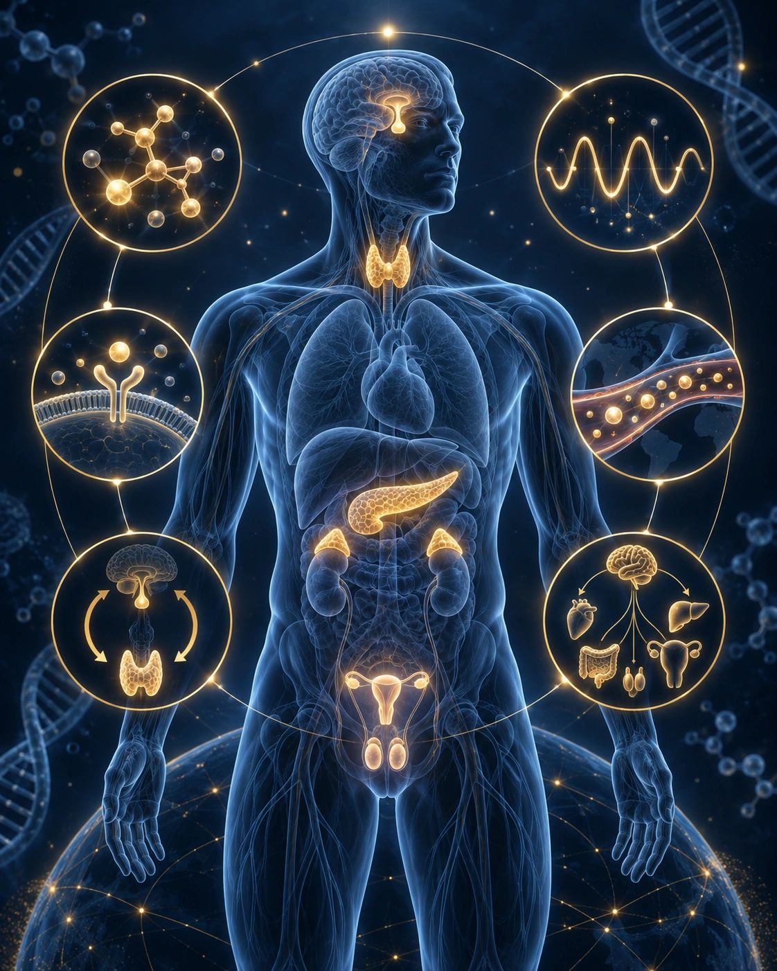
WHAT IS KPV?

KPV

IS KPV A MELANOCORTIN PEPTIDE?

THE IDENTITY MODEL

KPV stands for its amino-acid sequence:

LYSINE — PROLINE — VALINE

or:

Lys-Pro-Val.


It corresponds to the C-terminal sequence of:

α-MSH.


α-MSH itself is a:

13-AMINO-ACID PEPTIDE


derived from:

PROOPIOMELANOCORTIN — POMC.


That precursor also participates in the generation of several biologically important peptides.

THE IDENTITY MODEL

IS KPV A MELANOCORTIN PEPTIDE?

THE IDENTITY MODEL

POMC

↓

Peptide processing

↓

α-MSH

↓

C-terminal fragment

↓

LYS — PRO — VAL

↓

KPV

↓

Potential effects on:

INFLAMMATORY SIGNALING

CYTOKINE ACTIVITY

IMMUNE-CELL MIGRATION

BARRIER PHYSIOLOGY

INNATE HOST DEFENSE

↓

Potential:

CONTROLLED INFLAMMATORY RESPONSE


Again:

KPV is not simply:

A SMALLER α-MSH.


Fragmentation can preserve some biological functions while removing others.

That is an important concept throughout peptide pharmacology.

IS KPV A MELANOCORTIN PEPTIDE?

IS KPV A MELANOCORTIN PEPTIDE?

IS KPV A MELANOCORTIN PEPTIDE?

It is:

DERIVED FROM A MELANOCORTIN PEPTIDE.

But the pharmacology is interesting.

Full-length α-MSH interacts with:

MELANOCORTIN RECEPTORS.

These include:

MC1R 

MC2R 

MC3R 

MC4R 

MC5R 


The classical melanocortin receptor-binding motif is located elsewhere within α-MSH.


KPV does not contain that complete conventional receptor-binding sequence.

Experimental work has therefore suggested that at least some KPV anti-inflammatory activity occurs through:

MELANOCORTIN-RECEPTOR-INDEPENDENT MECHANISMS.


A classic animal study found that KPV's anti-inflammatory activity persisted despite melanocortin-receptor blockade and proposed interference with IL-1β-related signaling rather than conventional melanocortin receptor activation. 


But the story may not be entirely receptor independent.


Experimental colitis research suggests that some KPV effects may also depend partly on the broader:

MC1R / MELANOCORTIN ENVIRONMENT.


So the current biological picture is more nuanced:

KPV MAY OPERATE THROUGH BOTH MELANOCORTIN-RELATED AND MELANOCORTIN-INDEPENDENT PATHWAYS, DEPENDING ON TISSUE AND CONTEXT. 


That is more interesting than forcing the peptide into:

ONE RECEPTOR → ONE RESPONSE.

HOW KPV MAY WORK

INFLAMMATORY CYTOKINE SIGNALING

IS KPV A MELANOCORTIN PEPTIDE?

The recurring mechanistic themes include:

inflammatory cytokine regulation 

NF-κB-related inflammatory signaling 

IL-1β-related biology 

immune-cell migration 

barrier inflammation 

antimicrobial effects 

epithelial signaling 


Its biology therefore sits at the intersection of:

INFLAMMATION

and:

BARRIER DEFENSE.

INFLAMMATORY CYTOKINE SIGNALING

INFLAMMATORY CYTOKINE SIGNALING

INFLAMMATORY CYTOKINE SIGNALING

Inflammatory cytokines are molecular signals that allow immune cells and tissues to coordinate:

THE INFLAMMATORY RESPONSE.


Examples include:

TNF-α 

IL-1β 

IL-6 

chemokines 


These are not:

BAD MOLECULES.

They are necessary.


But excessive or persistent cytokine activity can contribute to:

tissue damage 

inflammatory bowel disease 

dermatitis 

chronic inflammatory signaling 

immune-cell recruitment 

barrier breakdown 


KPV-related research suggests it can attenuate aspects of:

PROINFLAMMATORY SIGNALING.

THE CYTOKINE MODEL

INFLAMMATORY CYTOKINE SIGNALING

INFLAMMATORY CYTOKINE SIGNALING

TISSUE STRESS / IMMUNE TRIGGER

↓

INFLAMMATORY CYTOKINES

↓

IMMUNE-CELL RECRUITMENT

↓

INFLAMMATION

If appropriately regulated:

↓

THREAT CONTROL + REPAIR

If excessive:

↓

TISSUE DAMAGE + BARRIER DISRUPTION


KPV may influence:

THE AMPLIFICATION PHASE OF THIS RESPONSE.


Again:

not:

ZERO IMMUNITY.


But potentially:

LESS EXCESSIVE SIGNALING.

NF-κB SIGNALING

NF-κB SIGNALING

NF-κB SIGNALING

NF-κB is one of the major transcriptional regulators of:

INFLAMMATION.


When activated, NF-κB can increase expression of genes involved in:

cytokines 

chemokines 

immune-cell recruitment 

inflammatory enzymes 

host-defense responses 


α-MSH biology has long been associated with suppression of excessive NF-κB activation, and this inflammatory-regulation network provides part of the biological background for KPV research. 

THE NF-κB MODEL

NF-κB SIGNALING

NF-κB SIGNALING

 INJURY / PATHOGEN / DANGER SIGNAL

↓

NF-κB ACTIVATION

↓

INFLAMMATORY GENE EXPRESSION

↓

CYTOKINES + CHEMOKINES

↓

IMMUNE RESPONSE


KPV-related signaling may reduce:

EXCESSIVE AMPLIFICATION

without necessarily abolishing:

THE ENTIRE HOST-DEFENSE RESPONSE.


That distinction is important.

IL-1β SIGNALING

NF-κB SIGNALING

IMMUNE-CELL MIGRATION

 IL-1β is a powerful:

PROINFLAMMATORY CYTOKINE.


It participates in:

fever 

leukocyte recruitment 

inflammatory amplification 

tissue responses to infection and damage


Experimental KPV work has suggested that some of its effects may involve:

INTERFERENCE WITH IL-1β-RELATED ACTIVITY.


In animal inflammatory models, KPV reduced leukocyte accumulation even when classical melanocortin pathways appeared not to explain the effect. 


This provides an important clue:

KPV may retain a highly specific part of α-MSH inflammatory biology despite being:

ONLY THREE AMINO ACIDS LONG.

IMMUNE-CELL MIGRATION

GUT AND BARRIER BIOLOGY

IMMUNE-CELL MIGRATION

Inflammatory tissues recruit:

neutrophils 

monocytes 

macrophages 

lymphocytes 


This movement is necessary during:

INFECTION AND INJURY.


But excessive cellular infiltration contributes to:

TISSUE DAMAGE.


KPV and α-MSH-derived peptides have shown experimental effects involving reduced:

INFLAMMATORY CELL MIGRATION AND ACCUMULATION. 


Again:

the biological goal may be:

APPROPRIATE CELL TRAFFICKING

rather than:

NO IMMUNE CELLS.

GUT AND BARRIER BIOLOGY

GUT AND BARRIER BIOLOGY

GUT AND BARRIER BIOLOGY

This is one of the most compelling research areas for KPV.


The gastrointestinal tract has an unusual problem.

It must tolerate:

FOOD

COMMENSAL MICROORGANISMS

and:

FOREIGN ANTIGENS

while simultaneously remaining able to detect:

PATHOGENS.


That requires extremely precise:

IMMUNE TOLERANCE + HOST DEFENSE.


Too little response:

↓

infection.

Too much response:

↓

inflammation and tissue injury.

THE BARRIER MODEL

GUT AND BARRIER BIOLOGY

GUT AND BARRIER BIOLOGY

INTESTINAL LUMEN

↓

EPITHELIAL BARRIER

↓

MUCOSAL IMMUNE SYSTEM

↓

Decision:

TOLERATE

or:

DEFEND


KPV is interesting because its parent melanocortin pathway participates in:

MUCOSAL IMMUNE REGULATION.

EXPERIMENTAL COLITIS

EPITHELIAL CELLS ARE IMMUNE CELLS TOO — FUNCTIONALLY SPEAKING

EPITHELIAL CELLS ARE IMMUNE CELLS TOO — FUNCTIONALLY SPEAKING

KPV has repeatedly been studied in:

ANIMAL MODELS OF COLITIS.


Reported effects have included reductions in:

inflammatory infiltration 

edema 

tissue injury 

weight loss 

histologic disease severity 


Reviews of melanocortin signaling in inflammatory bowel disease describe improvements across several experimental colitis models and suggest both MC1R-dependent and independent pathways may contribute. 


That makes KPV particularly interesting for:

GUT IMMUNOLOGY.


But the important concept is broader than:

“KPV FOR COLITIS.”


It is:

KPV MAY HELP MODULATE THE INTERFACE BETWEEN EPITHELIAL BARRIER FUNCTION AND INFLAMMATORY SIGNALING.

EPITHELIAL CELLS ARE IMMUNE CELLS TOO — FUNCTIONALLY SPEAKING

EPITHELIAL CELLS ARE IMMUNE CELLS TOO — FUNCTIONALLY SPEAKING

EPITHELIAL CELLS ARE IMMUNE CELLS TOO — FUNCTIONALLY SPEAKING

Epithelial cells are not just:

BRICKS IN A WALL.


They detect signals.


Produce cytokines.


Interact with microorganisms.


Communicate with immune cells.


Regulate permeability.


That means gut-barrier therapy cannot simply focus on:

TIGHT JUNCTIONS.


The epithelial surface is:

AN IMMUNOLOGIC ORGAN.


KPV becomes interesting precisely because:

BARRIER FUNCTION AND INFLAMMATION ARE CONNECTED.

ANTIMICROBIAL ACTIVITY

EPITHELIAL CELLS ARE IMMUNE CELLS TOO — FUNCTIONALLY SPEAKING

ANTIMICROBIAL ACTIVITY

This is one of the more unusual aspects of KPV.


Despite its inflammation-modulating effects, α-MSH and KPV have also demonstrated:

DIRECT ANTIMICROBIAL ACTIVITY

against organisms in experimental settings, including:

Staphylococcus aureus 

Candida albicans 


This is important because it challenges the simplistic model:

ANTI-INFLAMMATORY = WEAKER HOST DEFENSE.


The melanocortin system may help regulate inflammation while still contributing to:

INNATE DEFENSE. 


Again:

immune regulation and antimicrobial activity can coexist.

THE HOST-DEFENSE MODEL

THE HOST-DEFENSE MODEL

ANTIMICROBIAL ACTIVITY

PATHOGEN EXPOSURE

↓

Need:

MICROBIAL CONTROL

but also:

LIMITED COLLATERAL DAMAGE


KPV biology may potentially contribute to:

BOTH SIDES OF THAT EQUATION.


This is why:

IMMUNOMODULATION

is often a better word than:

IMMUNOSUPPRESSION.

SKIN BIOLOGY

THE HOST-DEFENSE MODEL

SKIN BIOLOGY

Skin presents a very similar challenge to the gut.


It is:

A BARRIER

and:

AN IMMUNE ORGAN.


Skin must manage:

microorganisms 

allergens 

UV exposure 

mechanical injury 

environmental chemicals 

while preventing:

CHRONIC INFLAMMATION.


α-MSH and related peptides have been extensively studied in models involving:

dermatitis 

cutaneous inflammation 

vasculitis 

fibrosis 


KPV is particularly interesting because it retains much of α-MSH's anti-inflammatory activity without the parent peptide's pigmentary effect.

DERMATITIS

THE HOST-DEFENSE MODEL

SKIN BIOLOGY

Inflammatory skin disorders often involve combinations of:

BARRIER DYSFUNCTION

IMMUNE ACTIVATION

ENVIRONMENTAL TRIGGER


KPV therefore has biologically plausible relevance to inflammatory skin research.


But again:

eczema.

contact dermatitis.

psoriasis.

rosacea.

infection.


They are not:

THE SAME DISEASE.


The inflammatory pattern matters.

THE α-MSH CONNECTION

PIGMENTATION VS INFLAMMATION

PIGMENTATION VS INFLAMMATION

To understand KPV fully, understand the parent peptide.


α-MSH is a:

NEUROIMMUNOMODULATORY PEPTIDE.


It helps connect:

NERVOUS SYSTEM

SKIN

IMMUNE SYSTEM

and:

ENDOCRINE SIGNALING.


This gives KPV an interesting heritage.

It is not simply:

A LAB-DESIGNED ANTI-INFLAMMATORY SEQUENCE.


It comes from a physiological system already involved in:

LIMITING EXCESSIVE HOST RESPONSE.

PIGMENTATION VS INFLAMMATION

PIGMENTATION VS INFLAMMATION

PIGMENTATION VS INFLAMMATION

Full-length α-MSH has:

MELANOGENIC ACTIVITY.


That means it can influence:

MELANIN / PIGMENTATION.


KPV lacks the full classical melanocortin pharmacophore necessary for those pigmentary effects.


Yet it retains important:

INFLAMMATORY-REGULATION ACTIVITY. 


This is a beautiful peptide-pharmacology example.

A larger peptide can contain:

MULTIPLE BIOLOGICAL MESSAGES.


Remove part of the molecule and you may retain:

ONE FUNCTION

while reducing:

ANOTHER.

THE CORE MECHANISM MAP

POMC

↓

α-MSH

↓

C-terminal:

KPV

↓

Potential modulation of:

NF-κB-RELATED SIGNALING

IL-1β-RELATED ACTIVITY

PROINFLAMMATORY CYTOKINES

IMMUNE-CELL MIGRATION

BARRIER INFLAMMATION

INNATE ANTIMICROBIAL DEFENSE

↓

Potential:

INFLAMMATORY CONTROL

BARRIER SUPPORT

HOST-DEFENSE COORDINATION


The core idea is:

  • KPV MAY HELP LIMIT DAMAGE WITHOUT SIMPLY TURNING IMMUNITY OFF.


WHAT THIS MECHANISM MAY SUGGEST

Mechanistically, KPV deserves research interest in:

  • inflammatory bowel disease 
  • intestinal barrier inflammation 
  • mucosal inflammatory disorders 
  • dermatitis 
  • inflammatory skin disease 
  • wound-associated inflammation 
  • selected allergic inflammatory states 
  • inflammatory tissue injury 
  • host-defense / barrier interactions 

Its strongest conceptual niche may be:

INFLAMMATION OCCURRING AT BARRIER SURFACES.

WHAT THIS MECHANISM DOES NOT PROVE

KPV biology does not prove that it:

  • treats every autoimmune disease 
  • cures inflammatory bowel disease 
  • eliminates gut permeability 
  • treats every infection 
  • replaces antimicrobial therapy 
  • should suppress every inflammatory response 
  • treats every rash 
  • is appropriate whenever inflammatory markers are elevated 

Again:

  • “INFLAMMATION” IS NOT A DIAGNOSIS.

Inflammation may be:

  • THE DRIVER

or:

  • THE RESPONSE TO THE DRIVER.

That distinction determines whether suppressing or modulating it is:

  • HELPFUL OR MISDIRECTED.

THE EVIDENCE, POTENTIAL APPLICATIONS, AND LIMITATIONS

 ENDOGENOUS / PHYSIOLOGICAL BIOLOGY

MEANINGFUL


KPV is directly derived from:

THE C-TERMINUS OF HUMAN α-MSH.


The parent melanocortin system has substantial established biology involving:

immune regulation 

inflammation 

skin 

central nervous system 

host defense 


A REAL PHYSIOLOGICAL IMMUNOREGULATORY SYSTEM.

PRECLINICAL EVIDENCE

MEANINGFUL

The strongest direct KPV evidence currently comes from:

  • cellular models 
  • inflammatory animal models 
  • experimental colitis 
  • skin/inflammation work 
  • antimicrobial experiments 

The research has repeatedly demonstrated biological activity despite the peptide's extremely small size.

HUMAN THERAPEUTIC EVIDENCE

EARLIER-STAGE

KPV's clinical therapeutic literature is considerably less developed than that of:

  • THYMOSIN ALPHA-1

or established therapeutic peptides such as:

  • GLP-1 ANALOGS.

But the underlying α-MSH / melanocortin inflammatory biology provides a substantial mechanistic foundation.

The appropriate conclusion is:

  • THERE IS A REAL BIOLOGICAL SIGNAL HERE.

The next translational question is:

  • HOW WELL DOES THAT SIGNAL PRODUCE CLINICALLY MEANINGFUL OUTCOMES IN SPECIFIC HUMAN DISEASE STATES?

That is the research question.

Not a reason to dismiss the biology.

Research

INFLAMMATORY BOWEL DISEASE

INFLAMMATORY BOWEL DISEASE

INFLAMMATORY BOWEL DISEASE

This may be KPV's most compelling research domain.


Experimental studies have examined KPV in:

DSS colitis 

immune-cell-transfer colitis 

other inflammatory bowel models 

Reported outcomes include improvements in:

weight 

inflammatory infiltration 

edema 

mucosal histology 

survival in selected models 


The melanocortin system itself is increasingly recognized as relevant to intestinal inflammatory regulation. 


Again:

the deeper concept is:

KPV MAY ALTER THE MUCOSAL IMMUNE ENVIRONMENT.

GUT-BARRIER DYSFUNCTION

INFLAMMATORY BOWEL DISEASE

INFLAMMATORY BOWEL DISEASE

KPV is frequently discussed in:

“LEAKY GUT.”

That phrase needs more precision.

Barrier dysfunction can occur in:

inflammatory bowel disease 

infection 

celiac disease 

medication injury 

severe systemic illness 

food-triggered inflammation 

metabolic dysfunction 


Again:

different cause.

Different therapy.


KPV may be most interesting where:

INFLAMMATORY SIGNALING ITSELF IS CONTRIBUTING TO BARRIER DAMAGE.


If the primary cause is:

ACTIVE CELIAC DISEASE WITH GLUTEN EXPOSURE,

a peptide does not replace:

REMOVAL OF THE DRIVER.

SKIN INFLAMMATION

INFLAMMATORY BOWEL DISEASE

CONTACT DERMATITIS AND ALLERGIC INFLAMMATION

KPV's derivation from α-MSH makes skin particularly relevant.


Melanocortin signaling participates in:

melanocyte biology 

keratinocyte signaling 

inflammatory regulation 

immune-cell behavior 


The ability of KPV to preserve anti-inflammatory signaling without strong pigmentary activity is one reason researchers have considered it attractive for inflammatory dermatologic applications. 


CONTACT DERMATITIS AND ALLERGIC INFLAMMATION

CONTACT DERMATITIS AND ALLERGIC INFLAMMATION

CONTACT DERMATITIS AND ALLERGIC INFLAMMATION

α-MSH-derived peptides have shown activity in experimental:

allergic contact dermatitis 

irritant dermatitis 

airway inflammation 

arthritis 


KPV may therefore have relevance across:

SELECTED IMMUNE-MEDIATED INFLAMMATORY ENVIRONMENTS. 


But:

allergy is not one pathway.


Again:

PATTERN MATTERS.

ANTIMICROBIAL RESEARCH

CONTACT DERMATITIS AND ALLERGIC INFLAMMATION

ANTIMICROBIAL RESEARCH

KPV's antimicrobial effects are especially intriguing because they suggest the peptide may:

LIMIT INFLAMMATION

without necessarily weakening:

DIRECT HOST DEFENSE.


Experimental research has reported activity against organisms including S. aureus and Candida albicans. 


That does not make KPV:

A GENERAL ANTIBIOTIC OR ANTIFUNGAL.


But it reinforces an important biological idea:

THE BEST IMMUNE RESPONSE IS NOT NECESSARILY THE MOST INFLAMMATORY RESPONSE.

FIBROSIS

CONTACT DERMATITIS AND ALLERGIC INFLAMMATION

ANTIMICROBIAL RESEARCH

Chronic inflammation can drive:

FIBROSIS.


Fibrosis represents excessive accumulation and remodeling of:

EXTRACELLULAR MATRIX.


Because melanocortin-related peptides can alter inflammatory and fibrotic signaling in experimental settings, KPV-related biology may have relevance to:

CHRONIC REMODELING STATES. 


Again:

organ fibrosis is complex.

Liver fibrosis.

lung fibrosis.

intestinal fibrosis.

skin fibrosis.

These are not interchangeable.

WHAT WE KNOW

WHAT WE THINK MAY BE HAPPENING

WHAT WE THINK MAY BE HAPPENING

KPV is a three-amino-acid peptide consisting of lysine, proline, and valine.


It is the C-terminal tripeptide of α-MSH.

Its parent peptide arises from POMC biology.


KPV retains substantial anti-inflammatory activity associated with α-MSH.


It does not retain the full classical melanocortin pharmacophore.


Its inflammatory effects are therefore not explained solely by conventional melanocortin-receptor activation.


IL-1β-related biology appears relevant.

NF-κB-related inflammatory regulation provides part of the broader α-MSH mechanistic context.


KPV can reduce inflammatory-cell migration in experimental models.


It has meaningful experimental evidence in colitis.


It has relevance to skin and barrier-tissue inflammation.


KPV and α-MSH have demonstrated antimicrobial activity in experimental systems.


Its biology is better described as immunomodulatory than simply immunosuppressive.

WHAT WE THINK MAY BE HAPPENING

WHAT WE THINK MAY BE HAPPENING

WHAT WE THINK MAY BE HAPPENING

KPV may function as:

A COMPACT INFLAMMATORY BRAKE DERIVED FROM A LARGER ENDOGENOUS IMMUNE-REGULATORY SIGNAL.


The tissue experiences:

INJURY / PATHOGEN / IMMUNE TRIGGER

↓

Inflammation begins

↓

Cytokines rise

↓

Immune cells migrate

↓

The response should control the threat

Then:

RESOLVE


KPV may help influence:

THE TRANSITION FROM AMPLIFICATION TOWARD CONTROL.


That could explain why it appears relevant in:

GUT

SKIN

and:

OTHER BARRIER TISSUES

where constant immune exposure makes:

OVERREACTION

particularly costly.


In that model:

KPV is not

TURNING OFF THE IMMUNE SYSTEM.


It may be helping the immune system:

STOP SHOUTING WHEN SHOUTING IS NO LONGER USEFUL.

WHAT REMAINS CONTEXT-DEPENDENT

WHAT WE THINK MAY BE HAPPENING

WHAT REMAINS CONTEXT-DEPENDENT

Important questions include:

Which inflammatory phenotypes respond best? 

What cellular target accounts for its receptor-independent effects? 

How much activity depends on MC1R or other melanocortin context? 

Which route produces useful tissue exposure? 

Is local delivery more biologically useful than systemic exposure for barrier disorders? 

What dose-response relationship exists in humans? 

Which inflammatory bowel phenotypes are most relevant? 

How does KPV interact with the intestinal microbiome? 

Can antimicrobial effects contribute meaningfully in vivo? 

Does prolonged inflammatory modulation alter host defense? 

How well does skin-model biology translate to different dermatologic diseases? 

Which inflammatory markers best reflect meaningful response? 


These are exactly the kinds of questions:

TRANSLATIONAL PEPTIDE MEDICINE SHOULD BE ASKING.

THE PRAXIS LENS

DO NOT BEGIN WITH KPV

Begin with:  WHY IS THIS TISSUE INFLAMED?   

Then:

PHYSIOLOGY

↓

PATTERN

↓

MECHANISM

↓

EVIDENCE

↓

RISK

↓

MONITORING

↓

DECISION

PHYSIOLOGY

Normal inflammation should:

RECOGNIZE

↓

RESPOND

↓

CONTROL

↓

RESOLVE

The problem may occur at any point.

Too little recognition:

  • INFECTION PERSISTS.

Too much response:

  • TISSUE DAMAGE.

Failure to resolve:

  • CHRONIC INFLAMMATION.

KPV appears most interesting around:

  • CONTROL + RESOLUTION.

EVIDENCE

Ask:

  • WHAT EVIDENCE SUPPORTS KPV IN THIS PARTICULAR TISSUE AND DISEASE MODEL?
  • Colitis?
  • Skin?
  • Antimicrobial biology?
  • General systemic inflammation?

Again:

do not take evidence from

  • ONE BARRIER SYSTEM

and automatically assume it applies to:

  • EVERY INFLAMMATORY CONDITION.

PATTERN

What is actually present?

  • inflammatory bowel disease? 
  • post-infectious inflammation? 
  • dermatitis? 
  • allergic inflammation? 
  • chronic barrier irritation? 
  • active infection? 
  • autoimmune activity? 
  • unexplained inflammatory marker elevation? 

Again:

  • THE WORD “INFLAMMATION” DOES NOT TELL US WHY THE PATIENT IS INFLAMED.

RISK

Potential considerations include:

  • inappropriate inflammatory suppression during active infection 
  • delayed identification of underlying disease 
  • allergic or local reactions 
  • product-quality problems 
  • route-related complications 
  • suppressing a signal that is still biologically necessary 

Again:

the issue is not

  • INFLAMMATION = BAD.

It is:

  • IS THIS PARTICULAR INFLAMMATION STILL SERVING A PURPOSE?

MECHANISM

Does KPV biology match:

  • THE DOMINANT BOTTLENECK?

If intestinal inflammation is driven by:

  • ONGOING PATHOGEN EXPOSURE,

the first question is:

  • WHAT IS THE PATHOGEN?

If dermatitis is driven by:

  • CONTINUED ALLERGEN EXPOSURE,

remove the:

  • TRIGGER.

If bowel inflammation is driven by:

  • UNTREATED CELIAC DISEASE,

address:

  • GLUTEN EXPOSURE.

If the driver is corrected but inflammatory signaling remains:

  • DISPROPORTIONATE OR NONRESOLVING,

then an immunoregulatory pathway becomes:

  • MORE BIOLOGICALLY RELEVANT.

MONITORING

Monitoring should follow:

THE TARGET TISSUE.

GUT

Potential outcomes:

  • stool frequency 
  • urgency 
  • abdominal pain 
  • bleeding 
  • mucosal disease activity 
  • disease-specific inflammatory markers when clinically relevant 
  • nutritional status 

SKIN

Potential outcomes:

  • erythema 
  • lesion burden 
  • itching 
  • barrier integrity 
  • flare frequency 
  • standardized photography 

HOST DEFENSE

Watch:

  • infection frequency 
  • infectious symptoms 
  • worsening local infection 
  • healing 

Again:

monitor

  • THE CLINICAL PATTERN.

Not simply:

  • A GENERIC “INFLAMMATION SCORE.”

THE PRAXIS 5

MECHANISM

KPV appears to influence:

  • INFLAMMATORY AMPLIFICATION + CYTOKINE SIGNALING + IMMUNE-CELL MIGRATION + BARRIER IMMUNITY.

Its α-MSH origin provides an important physiological foundation.

EVIDENCE

Experimental inflammatory evidence:

  • MEANINGFUL.

Gut / colitis biology:

  • PARTICULARLY INTERESTING.

Human therapeutic translation:

  • EARLIER-STAGE.

Again:

the evidence should guide:

  • THE CLAIM

without erasing:

  • THE BIOLOGY.

RISK

Do not confuse:

  • IMMUNOREGULATION

with:

  • UNIVERSAL IMMUNE SUPPRESSION.

And do not assume:

  • LESS INFLAMMATION IS ALWAYS BETTER.

The inflammatory response has a purpose.

MONITORING

Monitor:

  • THE DISEASE / BARRIER / FUNCTIONAL OUTCOME.

If KPV is being considered for gut biology:

  • monitor the gut.

If skin:

  • monitor the skin.

Again:

  • THE MECHANISM MUST CONNECT TO AN OUTCOME.

PATIENT CONTEXT

A patient with:

  • QUIESCENT IBD AND RESIDUAL BARRIER DYSFUNCTION

is not identical to:

a patient with:

  • SEVERE ACTIVE INFECTIOUS COLITIS.

A patient with:

  • ECZEMA

is not identical to:

someone with:

  • CELLULITIS.

A patient with:

  • POST-INFECTIOUS INFLAMMATION

is not the same as:

someone with:

  • AN UNCONTROLLED ACTIVE PATHOGEN.

Same peptide.

Completely different:

  • IMMUNE ENVIRONMENT.

SEVEN QUESTIONS TO ASK BEFORE KPV

1. WHAT INFLAMMATORY PROBLEM ARE WE ACTUALLY TRYING TO CHANGE?

  • gut? 
  • skin? 
  • mucosal? 
  • allergic? 
  • something else? 

2. WHY IS THE TISSUE INFLAMED?

  • infection? 
  • autoimmunity? 
  • barrier injury? 
  • allergen? 
  • mechanical damage? 
  • unresolved inflammatory signaling? 

3. DOES KPV'S BIOLOGY MATCH THAT MECHANISM?

  • cytokine regulation? 
  • IL-1β? 
  • immune-cell migration? 
  • mucosal inflammation? 
  • barrier signaling? 

4. WHAT TYPE OF EVIDENCE SUPPORTS THIS PARTICULAR USE?

  • cellular? 
  • animal? 
  • colitis model? 
  • dermatologic model? 
  • human? 

5. COULD REDUCING THE INFLAMMATORY SIGNAL INTERFERE WITH SOMETHING THE BODY STILL NEEDS TO DO?

  • fight infection? 
  • clear damaged tissue? 
  • heal a wound? 

6. WHAT WILL WE MONITOR?

  • symptoms? 
  • stool? 
  • skin? 
  • inflammatory activity? 
  • infection? 

7. WHAT WOULD DEFINE SUCCESS?

Not:

  • “INFLAMMATION WENT DOWN.”

But:

  • THE TISSUE FUNCTIONED BETTER WITHOUT COMPROMISING THE RESPONSE IT STILL NEEDED.

KPV

KEY TAKEAWAYS

  • KPV is a tripeptide composed of lysine, proline, and valine.
  • It is the C-terminal tripeptide of α-MSH.
  • α-MSH is derived from POMC and participates in neuroimmune and melanocortin signaling.
  • KPV retains important anti-inflammatory effects of α-MSH.
  • KPV does not contain the complete classical melanocortin receptor-binding motif.
  • Some of its inflammatory-regulation effects therefore appear to occur independently of traditional melanocortin receptor activation. 
  • The mechanism may not be entirely receptor independent in every tissue.
  • Experimental colitis research suggests that MC1R biology may still influence some KPV effects. 
  • KPV has been linked to regulation of inflammatory cytokine signaling.
  • IL-1β-related biology appears particularly interesting.
  • The broader α-MSH system regulates NF-κB-related inflammatory signaling.
  • KPV can reduce inflammatory-cell accumulation in experimental models.
  • Gut and barrier biology represent major research areas for KPV.
  • Experimental colitis studies have shown reductions in inflammatory and histological disease features.
  • Skin is another biologically relevant barrier system.
  • KPV preserves inflammatory-regulation activity without the strong pigmentary signaling of full-length α-MSH. 
  • KPV and related melanocortin peptides have demonstrated antimicrobial effects in experimental systems.
  • This suggests inflammatory regulation and host defense do not have to be opposites. 
  • KPV is better described as immunomodulatory than simply immunosuppressive.
  • The goal of inflammation is not zero inflammation.
  • The goal is an appropriate response followed by appropriate resolution.
  • KPV may be particularly relevant when the inflammatory response becomes excessive or fails to resolve.
  • Gut-barrier dysfunction should not automatically be interpreted as an indication for KPV.
  • The underlying driver still matters.
  • Active infection, celiac disease, structural disease, allergen exposure, autoimmune disease, medication injury, and other causes require different strategies.
  • The same principle applies to skin inflammation.
  • KPV should not be reduced to a universal “anti-inflammatory peptide.”
  • Its most useful biological identity is an α-MSH-derived inflammatory-regulation and barrier-signaling peptide.
  • The best question is not “does KPV lower inflammation?”
  • The better question is “why is this tissue inflamed, does the inflammatory response remain necessary, has it become disproportionate or nonresolving, does KPV biology intersect with that bottleneck, and what measurable improvement would demonstrate restoration of barrier or tissue function?”

THE BOTTOM LINE

KPV is a perfect example of why peptide medicine should not be judged by:

  • MOLECULE SIZE.

Three amino acids:

  • LYSINE
  • PROLINE
  • VALINE

Yet those three amino acids preserve a meaningful portion of the inflammatory-regulation biology of:

  • α-MSH.

That matters because α-MSH belongs to one of the body's most interesting communication systems:

  • THE MELANOCORTIN SYSTEM.

A system connecting:

  • BRAIN
  • IMMUNE CELLS
  • SKIN
  • BARRIER TISSUES
  • INFLAMMATORY CONTROL.

KPV appears to extract a particularly interesting biological message from that larger peptide:

  • CONTROL THE RESPONSE WITHOUT NECESSARILY SHUTTING DOWN DEFENSE.

That makes it relevant to places where the body faces the hardest immunological balancing act.

Especially:

  • THE GUT
  • and:
  • THE SKIN.

Both must constantly decide:

  • WHAT SHOULD I TOLERATE?
  • and:
  • WHAT SHOULD I ATTACK?

Too little defense:

infection.

Too much defense:

tissue injury.

That is why KPV should not simply be described as:

  • AN ANTI-INFLAMMATORY PEPTIDE.

The deeper idea is:

  • INFLAMMATORY PROPORTIONALITY.

Can the immune system mount enough inflammation to:

  • CONTROL THE THREAT

without creating enough inflammation to:

  • DESTROY THE BARRIER IT IS TRYING TO PROTECT?

KPV may participate in that balance.

And that changes the clinical question.

Instead of:

  • “THE PATIENT HAS INFLAMMATION — SHOULD WE USE KPV?”

ask:

  • “WHAT TRIGGERED THE INFLAMMATION, IS THE TRIGGER STILL PRESENT, IS THE RESPONSE APPROPRIATE TO THAT TRIGGER, HAS THE SYSTEM FAILED TO RESOLVE, DOES KPV'S α-MSH-DERIVED BIOLOGY MATCH THE DYSREGULATED NODE, AND WHAT functional outcome would show that the tissue became healthier rather than merely less inflamed?”

That is the difference between:

  • SUPPRESSING INFLAMMATION

and:

  • UNDERSTANDING IMMUNE REGULATION.


STOP MEMORIZING PEPTIDES. START UNDERSTANDING THE SYSTEM.

  Praxis Peptide Institute provides educational information and is not a substitute for individualized medical diagnosis or treatment. KPV is an α-MSH-derived tripeptide with experimental activity involving inflammatory regulation, barrier biology, immune-cell migration, and host-defense signaling. Its relevance may differ substantially according to the underlying cause of inflammation, presence of infection, autoimmune disease, barrier injury, medication exposure, tissue involved, route of administration, concurrent therapy, and therapeutic goal. Decisions involving KPV or other immunoregulatory peptides should focus on the biological driver of inflammation, the evidence applicable to the particular tissue and condition, product quality, meaningful disease or functional outcomes, and whether modulation of the inflammatory response is appropriate at that stage of illness. 

Copyright © 2026 Praxis Peptide Institute - All Rights Reserved.

Powered by

This website uses cookies.

We use cookies to analyze website traffic and optimize your website experience. By accepting our use of cookies, your data will be aggregated with all other user data.

Accept